WHAT IS THERMAL IMAGING? 
Thermal Imaging is an effective, safe, non-invasive tool for diagnosing areas of inflammation or injury by measuring differences in surface temperature of the animal. It also gives special insights into soft tissue inflammation that is occurring as a result of compensating for a primary injury. In addition, thermal imaging is an excellent tool for monitoring the healing progress of soft tissue or joint. Thermal images can depict areas of heat, inflammation, cold, and reduced blood flow in the muscular, vascular, skeletal and nervous systems.
HOW DOES THERMAL IMAGING WORK?
Thermal imaging utilizes a specialized camera that converts emitted heat or infrared waves into a picture visible to the human eye. Thermal Imaging is a physiologic imaging tool. This means it tell us what is going on right now in terms of changes in blood flow. When an animal has inflammation or injury to an area, changes in blood flow occur. These are seen as changes of color on the image of that area.
WHAT ARE SOME OF THE USES OF THERMOGRAPHY?
Thermal imaging can be used to help identify:
- Muscular pain and injury
- Back problems and injury
- Joint and skeletal issues
- Tendon and ligament issues
- Identification of lameness and gait abnormalities
- Hoof balance
- Hoof abscesses and bruises
- Establishment of a baseline to track changes over time
Examples of equine thermal images showing areas of increased heat (white & red emit the most heat):


PREPARING YOUR HORSE FOR EQUINE THERMOGRAPHY:
As equine thermographs can be affected by artifacts (anomalies in the image), guidelines should be followed to ensure the most accurate and easily interpreted images are taken.
1. Your horse must be mud-free and completely dry. Mud and water block the emission of infrared and affect temperature measurements.
2. Better views of the neck are obtained where the mane is braided or put into bunches along the crest at least 30 minutes before the session.
3. Your horse should be stabled for an hour before the consultation, unless stabling causes them to be agitated or stressed. Weaving or door banging will make the images of the front legs misleading. If this is the case then the horse should remain turned out.
4. Your horse should be groomed, and feet picked out, but don’t groom the horse within 20 minutes prior to the consultation time. Grooming increase blood flow to the area. Don’t pull the mane or tail within 24 hours prior to the consultation.
5. Take off all bandages and boots 20 minutes prior to the consultation time; otherwise the horse’s legs will appear to be warmer than they are. Take off all blankets. If the horse must wear one, try to limit it to a cotton summer sheet and fasten loosely.
6. Don’t use any coat conditions, fly sprays, blister, liniments, poultices, creams, and so forth on the day of the consultation, unless under veterinary direction.
7. Make a note of any medications the horse is taking.
8. Prepare an area suitable for imaging. This should be out of direct sunlight (the radiation from the sun will warm your horse and create falsely warm areas), and drafts (the breeze will “take away” infrared radiation coming from your horse and make the area appear cooler).
9. Ideal areas are a closed barn or sheltered area of the yard with a level hard surface. Images can be taken in less than perfect conditions, but the resulting report won’t be as easily to interpret.
10. Finally, if the temperature is greater than 25%, the horse will naturally appear warmer as hiss capillary beds dilate and blood flow increases to cool him down. On hot summer days it is best to take images as early as possible in the morning, while the horse is still cool, or late in the evening. Try to avoid midday. External temperatures are always noted when interpreting images.
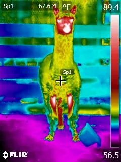
A few more examples of different animals being thermal imaged. You can note the different heat patterns.


PREPARING YOUR DOG OR CAT FOR THERMOGRAPHY:
Dogs & cats should not be bathed, go swimming, sit in front of a heater or air conditioner, sit in a window sunspot, or come in wearing harnesses, booties or vests (unless the goal is to evaluate the fit of that item).
Examples of Canine Thermal Images:


Veterinary thermal imaging fills a gap in clinical diagnostic tools and helps show the animal’s physiological state by graphically mapping skin surface temperature in response to changes in blood flow. In healthy animals, the thermal pattern on the skin is symmetrical. This is because skin blood flow is controlled by the sympathetic nervous system.